Probleemstelling:
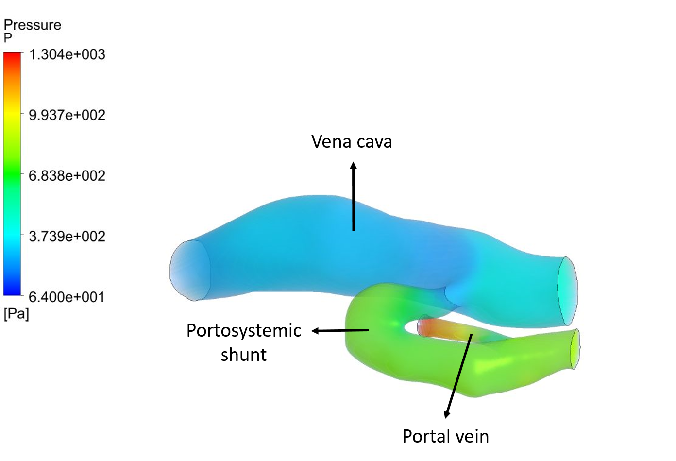
The portal vein is a very important blood vessel in which all nutrient- and toxin-rich blood is collected that comes from the gastro-intestinal organs. The portal vein drains into the liver who detoxifies the blood before it enters the systemic circulation (Figure 1). A portosystemic shunt is an anomalous vein that allows portal blood to enter the systemic circulation without first passing through the liver (Figure 2). This is a congenital problem widely described in dogs. Abdominal ultrasound and/or computed tomography are the medical imaging techniques of choice to confirm the presence and type of portosystemic shunt. They are interpreted in a rather static way, not revealing any information on the hemodynamics.
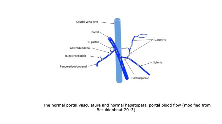
Figure 1. The normal portal vasculature and normal hepatopetal portal blood flow (blood flow towards the liver) (modified from Bezuidenhout 2013)
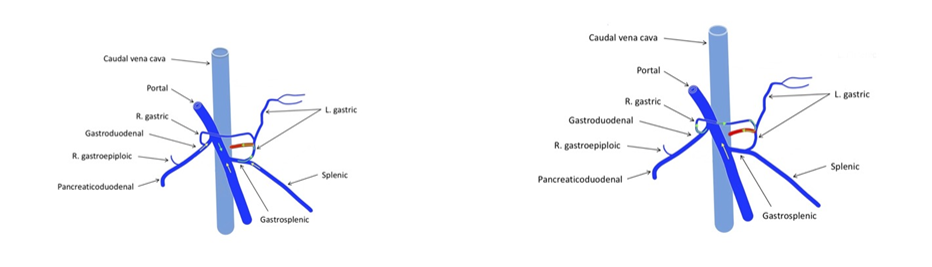
Figure 2. The portal vasculature in the presence of a portosystemic shunt and two possibilities of hepatofugal portal blood flow (blood flow away from the liver)
Gradual attenuation of the shunt is intended to redirect the blood through the portal branches, which are often hypoplastic and thus need time to adapt to the increased blood flow. The surgical approaches currently used result in incomplete closure of the shunt in 15-30% of the patients. Some of the dogs with a partially attenuated congenital portosystemic shunt have excellent clinical outcomes, whereas others have poor outcomes.
It has been hypothesized that both the blood flow direction (hepatopetal versus hepatofugal) in the segment of the portal vein cranial to the origin of the shunt as well as in the portosystemic shunt adjacent to the portal vein might indicate whether re-operation to close the portosystemic shunt completely should be advised or not in dogs with a patent portosystemic shunt after the first surgery.