Probleemstelling:
Zebrafish are more and more being used in cardiovascular research to study the impact of genetic disease, assess the effect of drugs on physiology and development or for precision medicine where a patient-tailored zebrafish model is created to test the efficacy of drugs.
While the biology and genetics of the zebrafish are well understood, this is much less the case for its hemodynamics and biomechanical forces acting on the cardiovascular tissues. Despite that the circulation of the fish is fundamentally different from the mammalian circulation, blood flow is pulsatile and it seems that pressure pulsations are damped by a peculiar trumpet-like structure (the bulbus arteriosus) to ensure steady flow through most of the circulation. A second peculiarity is the fact that, due to the small dimensions of the vessels, blood can no longer be considered as a homogeneous liquid, and blood flow should be addressed as a multi-phase flow, impacting the shear forces that are acting on the arterial wall.
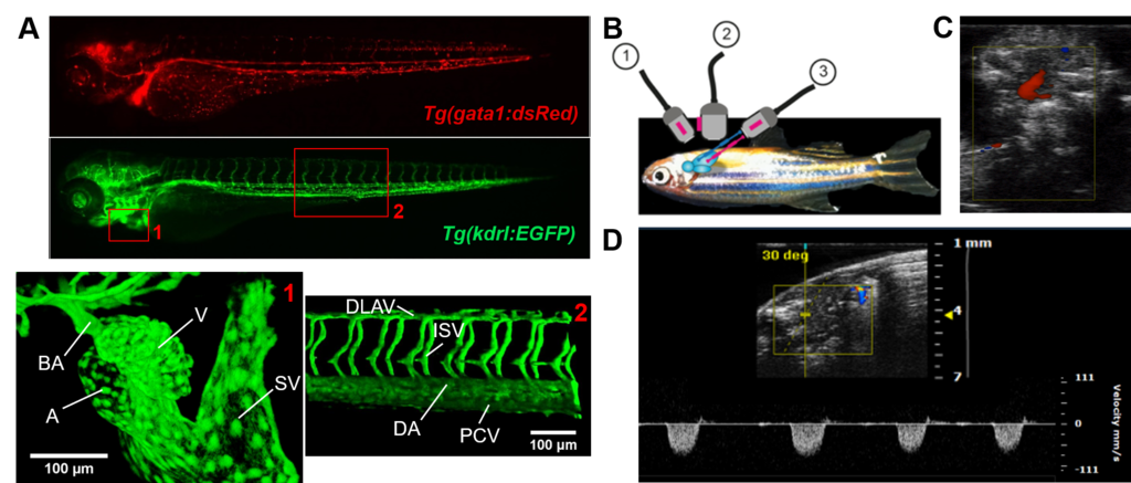
Figure 1: Imaging of the cardiovascular system in zebrafish larvae and adults. A) Top: epifluorescent images of 3-day old Tg(gata1:dsRed) and Tg(kdrl:EGFP) transgenic zebrafish larvae. The gata1 promotor is active in erythrocytes (similar to the globin promotor proposed in this project), leading to red fluorescent labeling of circulating cells. The kdrl:EGFP line expresses the EGFP protein specifically in endothelial cells, labeling blood vessels and the endocardium. Bottom: 3D reconstructions of 2-photon confocal images recorded at the indicated anatomical sites. A: atrium, BA: bulbus arteriosus, SV: sinus venosus, V: ventricle, DA: dorsal aorta, DLAV: dorsal longitudinal anastomotic vessel, ISV: intersegmental vessel, PCV: posterior caudal vein. Images were recorded by Dr. Sips at the Brigham and Women’s Hospital, Boston. B) Schematic overview of adult zebrafish positioned for ultrasound imaging, with three defined transducer positions illustrated (1: Short Axis (SAX), 2: Long Axis (LAX), 3: Abdominal-Cranial Axis (ACX)). Adapted from (11) C) Color Doppler imaging of cardiac inflow (ACX). D) Pulsed-wave doppler imaging of cardiac outflow (LAX). Ultrasound images were recorded using the Visualsonics 2100 platform at the Infinity small animal imaging core facility, UGent.