32375 MR elastography of the brain - a way to measure intracranial pressure? A computational/experimental study
Begeleider(s): ir. Sarah Vandenbulcke

Richtingen: Master of Science in Biomedical Engineering

Probleemstelling:

Intracranial pressure is elevated in patients with traumatic brain injury and in neurological disorders as hydrocephalus or Chiari malformation. It is a vital physiological parameter which, unfortunately, cannot be assessed in a non-invasive way. However, given the poroelastic nature of the brain parenchyma and its mechanical confinement within the skull, we hypothesize that theer is a coupling between the interstitial pressure in the brain and the functional stiffness of the tissue. The latter can be measured using Magnetic resonance elastography (MRE), a non-invasive tool to infer the stiffness and viscoelastic properties of the brain and spinal cord from the propagation of mechanically induced shear waves. Our hypothesis is supported by a recent study, where MRE during Valsalva (elevated intrathoracic pressures, known to lead to an increased intracranial pressure) showed transiently elevated brain stiffness.  

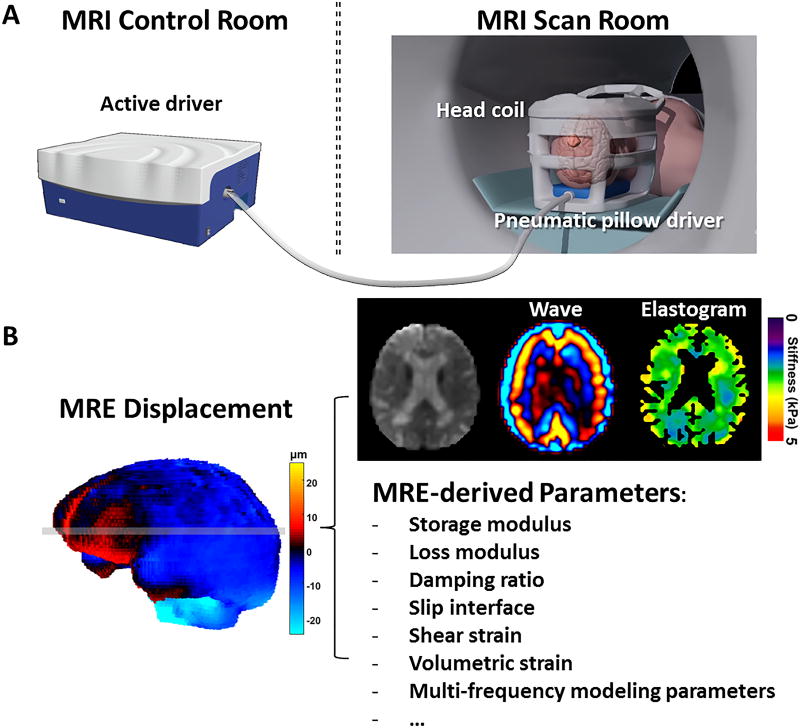
MRE involves 3 steps: (i) mechanical actuation to induce shear waves using an internal (arterial pulsations) or external vibrating source; (ii) imaging tissue displacements, and (iii) converting tissue displacements into stiffness properties (an elastogram) using an inversion algorithm. Inversion algorithms assume underlying isotropic linear elastic/viscoelastic constitutive material properties. In this thesis, the main focus is on step (iii), where we want to expand our experience in experimental and in-silico modelling of (ultrasound) shear wave elastography in complex anisotropic cardiovascular tissues to the brain and central nervous system.


Doelstelling:

In essence, we aim to use the 3D poroelastic computational and/or experimental models to study the propagation of shear waves under varying interstitial pressure conditions to allow us to assess how interstitial pressure relates to the functional stiffness of the brain and its manifestation in MRE.

Computational model: To get a solid understanding on the sensitivity of solutions to numerical settings (mesh dimensions, time steps and boundary conditions), we first model shear wave propagation in geometrically simplified beam-shaped volume of homogeneous, isotropic materials subjected to an intrinsic (breathing, cardiac pulsations) or external vibration source of varying frequencies (0.2 - 60 Hz). The next step is to model shear wave propagation in more advanced 3D brain models. We will in particular address (i) the impact of the vibration source (internal or external), location (for cranial/spinal assessment) and its frequency; (ii) isolated sustained and transient changes in CNP; (iii) isolated changes in solid stiffness; (iv) combined changes in CNP and stiffness; (v) simulate pathology. All modelling will be done in COMSOL Multiphysics, offering the advantage of built-in functions and the flexibility to implement dedicated material laws. Depending on the complexity of the problem at hand and required computational resources, simulations will run on stand-alone hardware or Ghent University high performance computing infrastructure.

Experimental model: In vitro experiments will be performed using the Siemens MAGNETOM Prisma 3.0 Tesla research scanner available within the Ghent Institute for functional and Metabolic Imaging (GIfMI). The scanner is equipped with the multi-cap system (THEA-Devices GmbH) that allows for multi-frequency shear wave activation using compressed air pads and GIfMI has a research agreement with the BIOQIC research programme of the Charité – Universitätsmedizin Berlin providing access to MRE sequences and inversion software, converting displacement data into stiffness maps. Phantoms of varying complexity will be designed and built, ranging from simple passive beam-shaped volumes of poroelastic tissue mimicking materials (with tofu an excellent candidate material [49]) to more realistic CNS phantom models that account for the brain parenchyma, CSF spaces and CSF flow, and allow dynamic arterial pulsations and changes in CNP due to breathing and blood volume shifts.

Depending on the interest(s) of the student(s), the topic will be further defined and narrowed down to a project apt for a master thesis.