Probleemstelling:
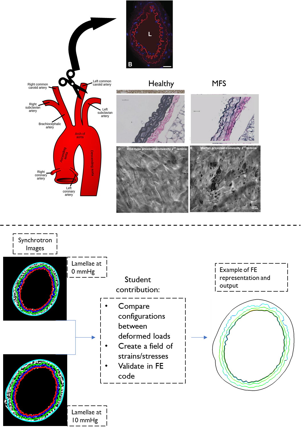
Marfan Syndrome (MFS), is characterized by disruptions of connective tissue that offer the structural foundation for cellular attachment and biomechanical load-bearing of soft tissues in the body. Elastin Lamellae, which are otherwise arranged as cylindrical layers of fenestrated elastin sheets coaxial to the direction of blood-flow, become fragmented with enlarged fenestrations in acute pathophysiology of MFS. A consequence, or concomitance, of these disruptions is the compromised biomechanical response, and the formations of aneurysms in the segments of the arterial tree which bear the highest loads, such as in the ascending portion of the thoracic aorta, or in the branching spot of the abdominal aorta. In more fatal occurrences, the inner walls of the aorta come apart and a dissection forms, which eventually leads to rupture of the aortic wall and internal bleeding followed by patient death.
In order to understand how the structural damage in elastin can lead to such pathology, a more thorough understanding of the interplay between elastin and the remaining matrix environment is needed. Our lab at BioMMedA has previously demonstrated experimental work that supports the hypothesis that lamellar damage occurs at locations of the highest stresses in mouse aortas, coinciding at regions of branching. Additionally, further experimental work has provided us with cross-sectional images of mouse carotid arteries under different loading conditions.
To that end, the goal of this project would be to gain additional insight on elastin biomechanics from these experimentally obtained images. The current framework for assessment of soft tissues on this scale uses the Finite strain theory to constitutively model the tissue as a homogeneous continuum made of different components. We seek to study how the configurations and changing conformations of these lamellae at different arterial loading can inform these models, and if they can accurately portray damage in our models.